

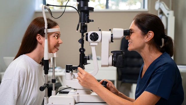
You go for a routine eye test to update your glasses, and your eye doctor spends a little longer than usual examining the back of your eye. Then comes the sentence you were not expecting: your optic nerve looks swollen. You feel fine, your vision is sharp, perhaps you have a mild headache. Yet within the hour, you are on your phone reading alarming articles about brain tumours.
That fear is entirely understandable. However, optic disc swelling is a clinical finding that demands a methodical explanation, and that explanation is frequently reassuring. If you or someone you care about has been told your optic nerves appear puffy, here is how we approach the evaluation step by step.
What the doctor sees: Anatomy in brief
All the nerve fibres from the light-sensitive retina converge to form the optic nerve, which bridges the eye directly to the brain. Leaving the back of the globe through the optic canal, its terminal head forms the circular optic disc.
Remarkably, the optic disc is the only portion of the central nervous system's wiring that a clinician can directly visualise through the pupil without invasive imaging. When that disc appears elevated, hazy, or blurred at the margins, we categorise the underlying causes into three primary clinical scenarios:
1. Raised Pressure Inside the Skull (Papilledema)
When intracranial pressure rises, it exerts hydrostatic pressure on the optic nerve sheaths, causing both optic discs to swell. Clinically termed papilledema, the most frequent driver I encounter in practice is Idiopathic Intracranial Hypertension (IIH). This is a condition predominantly affecting young women of childbearing age, often following recent weight gain.
- Classic Symptoms: Morning-predominant headaches, a pulsatile "whooshing" sound in the ears (tinnitus), and transient visual obscurations (momentary grey-outs when standing or bending over). Some patients experience double vision, while many others remain completely asymptomatic.
- Secondary Causes: Intracranial mass lesions, cerebral venous sinus thrombosis, hydrocephalus, malignant hypertension, and specific medications (such as tetracycline-class antibiotics and vitamin A derivatives).
- Clinical Imperative: Chronic elevation of intracranial pressure causes gradual axonal loss. Peripheral and side vision fade silently long before central acuity drops, which is why prompt investigation is vital.
2. Inflammation of the Nerve (Optic Neuritis)
Unlike papilledema, optic neuritis typically affects a single eye in young adults. Vision blurs over several days, colours appear desaturated (washed out), and eye movements provoke sharp discomfort. Interestingly, the optic disc appears swollen in only about one-third of cases because the inflammatory lesion frequently sits further back along the nerve, completely out of direct view (retroburbar neuritis).
Because optic neuritis can serve as an early sentinel sign of multiple sclerosis or antibody-mediated demyelinating spectrum disorders, arranging an immediate MRI and initiating timely therapy alters long-term neurological outcomes.
3. Pseudopapilledema: A Disc That Only Looks Swollen
Not all swollen-appearing nerves are diseased. Many individuals possess structurally crowded, small optic discs, particularly in hypermetropic (farsighted) eyes, that mimic true edema. Another common mimic is optic disc drusen, which are benign, calcified protein deposits buried within the nerve head that frequently run in families. Individuals with pseudopapilledema go through life with entirely normal vision and require no treatment. Distinguishing a harmless structural variant from true swelling requires specialised diagnostic testing; a specialist will never dismiss a swollen disc as "probably nothing" without objective confirmation.
How We Sort It Out: The Diagnostic Pathway
When faced with optic disc elevation, a definitive diagnosis relies on a structured protocol:
- Comprehensive Neuro-Ophthalmic Examination: Assessment of visual acuity, colour plates, pupillary reflexes, formal visual fields, and blood pressure measurement.
- Optical Coherence Tomography (OCT): A rapid, non-invasive scan that measures retinal nerve fibre layer thickness, quantifies disc elevation, and helps identify buried drusen.
- Advanced Neuroimaging: If true swelling is confirmed or suspected, an MRI of the brain and orbits with a magnetic resonance venogram (MRV) is mandatory to evaluate venous sinuses and rule out structural masses. Crucially, imaging always precedes a lumbar puncture to ensure a safe opening pressure measurement.
- Lumbar Puncture (Spinal Tap): Performed with the patient in a lateral decubitus position once imaging is clear. In adults, an opening pressure exceeding 25 mm of water confirms intracranial hypertension, and cerebrospinal fluid is sent for baseline laboratory analysis.
Red Flags: When to Seek Urgent Evaluation
Seek immediate evaluation at an emergency department or specialised eye centre if you experience:
- Transient blackouts or grey-outs of vision.
- Sudden onset double vision.
- Severe, unrelenting headaches accompanied by nausea or vomiting.
- Any acute drop in central or side vision in either eye.
If none of these acute warning signs are present, you should still go for a comprehensive neuro-ophthalmic evaluation within a few days rather than weeks. A timely, methodical assessment turns uncertainty into a clear plan, safeguarding your vision for the long term.
Disclaimer: The opinions expressed within this article are the personal opinions of the author. NDTV is not responsible for the accuracy, completeness, suitability, or validity of any information on this article. All information is provided on an as-is basis. The information, facts or opinions appearing in the article do not reflect the views of NDTV and NDTV does not assume any responsibility or liability for the same.