

)
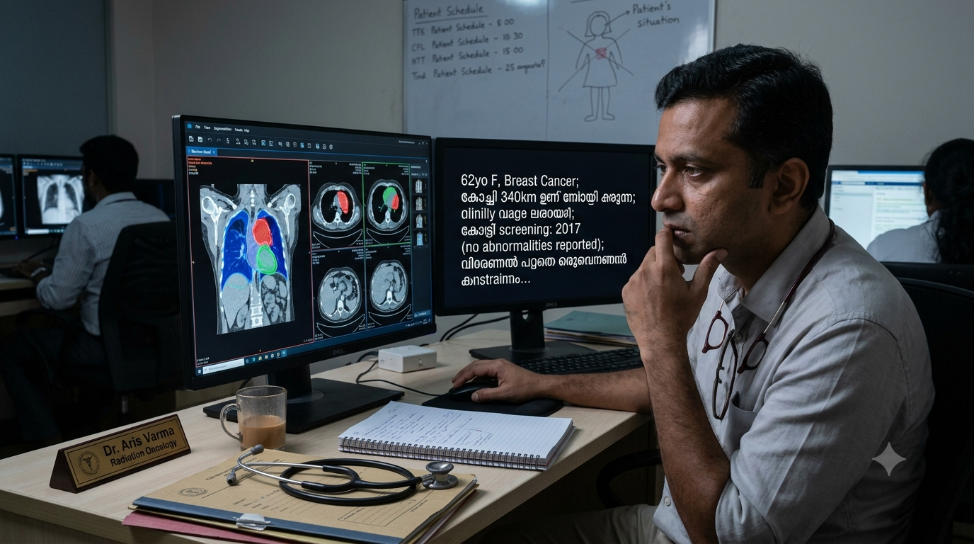
On the planning CT scan, a radiation oncologist has to outline the volume of tissue that will receive a radiation dose and the organs that have to be protected from it. This author does this unglamorous work on most days of the week, and it involves moving from slice to slice to outline the areas that will receive radiation and the organs that will be spared. This is where an imaging algorithm (built to automate this workflow) would probably meet a real treatment decision. Most algorithms fail to live up to the hype.
The "specificity" lure of the AI tools
Almost every AI product promises to read the image more accurately; at least the marketing brochure promises that. Any trained radiologist can accurately identify the lesions in the scan; the premise for automation is the lack of an adequate number of trained personnel. It was for that reason that the World Health Organization endorsed computer-aided detection software, which reads a chest film and flags the ones that look suspicious, for tuberculosis screening in 2021.
In a screening programme, the figure that determines cost is specificity, which is the proportion of healthy people a test correctly clears. A study of one widely used chest X-ray tool in a Lima hospital reported sensitivity of 0.91 and specificity of 0.32. Nine cases in ten were caught, and two thirds of the people who did not have tuberculosis were flagged. Each of these requires a sputum test and a repeat film, and, in a consequence more relevant for India, a day of lost wages. Nobody has published what that cascade costs per thousand people screened in an Indian district, and that is the figure a health secretary would want before signing anything.
The screening premise in India has also been imported without much examination. India runs no population-wide mammography programme, although national policy offers a clinical breast examination (a physical examination by a health worker) once every five years. The fifth National Family Health Survey found that under one per cent of women had ever received one. The women who die of breast cancer in this country are largely those who never entered the screening pathway, rather than those whose mammogram was misread. Hence, the "AI solution" appears redundant, and someone has to underwrite the cost of mammography.
These tools are cleared as aids to a human reader rather than as replacements, which means a doctor still signs the report and still carries the legal responsibility for it. That arrangement contains a circularity worth naming. If every AI output is checked by a person, the reading time the product was sold to save has not been saved. If the outputs are not checked, a clinician is signing something they did not verify.
The workload has shifted towards increasing Computed Tomography (CT) or Magnetic Resonance Imaging (MRI) scans, and the interpretative burden on the radiologist has increased manifold. Equivalent studies to measure the impact on trained radiologists are lacking in India. CT scans routinely turn up incidental findings, which are small unexplained abnormalities that nobody was looking for and that somebody then has to prove benign. Most of the AI products being sold are still built for the chest radiograph. The reason is commercial. Large public collections of labelled chest films are freely available, so a competent team can train a respectable model within months. Administrative clearance depends on whether the product is broadly equivalent to something already on the market. Commercial incentives align poorly with actual clinical need.
The cost of keeping the system running
The associated cost of product subscription, archived images, storage, reliable uptime and version management is the most understated aspect of "AI in healthcare". Who pays for it? The cost burden will eventually be transferred to consumers, something that they never signed up for. Even in the United States, payment for radiology algorithms remains fragmented and largely unresolved, with only a handful of tools securing dedicated reimbursement out of the hundreds cleared.
What the published evidence shows
Clinical evidence is more sober than marketing claims. A systematic review commissioned by the United Kingdom's screening committee found no prospective study of accuracy in real screening practice. Of the thirty-six systems tested against the original radiologist's decision, thirty-four were less accurate than a single radiologist and every one was less accurate than two radiologists reading together. This holds true for radiation oncology systems as well, although the evidence is sparse.
Questions worth asking before buying an AI system for a healthcare facility
There are five serious questions that no one is asking. Surprisingly, none of them requires technical training, only common sense:
1. Which set of patients formed the validation cohort for the AI product?
2. What was the tool measured against, human labels or something that actually happened to a patient?
3. What is its specificity at the threshold the hospital intends to use?
4. Who checks its output, and who answers for it when it is wrong?
5. Once integration, storage and support are included, what is the running cost?
The technology works well enough. The gains from it are uneven, and they are not yet reaching the patient. This opens up an opportunity to define the algorithms for the large pool of Indian patients, provided we are able to justify the returns on investment. Separate the hype from the marketing brochure and investors desks before clinical use is the most important takeaway.
(Dr. Abhishek Puri, Associate Consultant Radiation Oncology, Fortis Mohali)
Disclaimer: The opinions expressed within this article are the personal opinions of the author. NDTV is not responsible for the accuracy, completeness, suitability, or validity of any information on this article. All information is provided on an as-is basis. The information, facts or opinions appearing in the article do not reflect the views of NDTV and NDTV does not assume any responsibility or liability for the same.