

)
Cardiology and ophthalmology used to operate in separate silos. If your heart was at risk, you got cholesterol panels and treadmill tests. If your eyes needed attention, you got a vision check and an eye pressure reading. That separation is starting to blur. Research shows that your routine eye exam could become an early warning system for heart disease.
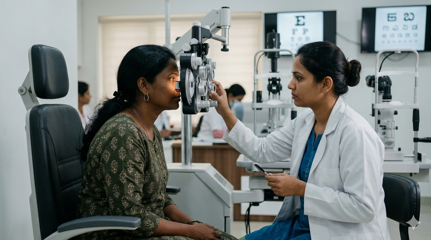
When an ophthalmologist examines your retina through a slit lamp or a high-resolution scan, glaucoma and macular degeneration aren't the only things on the checklist. The retina is the only place in the body where blood vessels can be seen directly and non-invasively, in real time, without an incision or a dye injection. And it shares structural and embryological origins with the coronary arteries feeding your heart.
Why The Retina Works As A Proxy
The retina is technically an extension of the brain, and its tiny arterioles and venules are a direct map of your microvasculature. When chronic hypertension, inflammation, or high cholesterol starts damaging arterial walls, it doesn't happen only in the heart. The same wear shows up in the back of the eye, often before it shows up anywhere else that's easy to check.
What Retina Scans Can Pick Up
When chronic issues like high blood pressure, inflammation, or high cholesterol start damaging your body, the wear and tear often shows up in the back of the eye first:
- Narrowing Blood Vessels: High blood pressure thickens the walls of retinal arteries, leaving less room for blood to flow. This arteriolar narrowing, whether generalised or in specific spots, is a direct signal of systemic vascular resistance.
- Crossing Changes (AV Nicking): Stiff, high-pressure arteries can push down on intersecting veins, causing them to bulge.
- Tiny Leaks: Small balloon-like spots or micro-haemorrhages signal that the inner lining of the blood vessels is breaking down, an early step toward hardening of the arteries.
- Cholesterol Plaques: Rarely, doctors spot a bright yellow cholesterol fragment (a Hollenhorst plaque) travelling toward the brain or heart, requiring an immediate cardiac workup.
From Reactive To Predictive
Cardiology has long relied on risk calculators built from age, smoking status, cholesterol, and blood pressure to estimate ten-year heart attack risk. These scores routinely miss people in the "intermediate risk" category: people whose numbers look fine but who go on to have sudden cardiac events.
Retinal imaging adds a physical layer to that picture. By analysing vessel width, tortuosity, and the branching pattern of the vascular tree, AI-assisted retinal analysis can estimate a patient's vascular age rather than just their calendar age. Large studies, including UK Biobank-based models, have shown retinal vascular scoring predicts stroke and heart attack risk about as well as standard risk calculators like Framingham.
Some of that research also found that adding retinal data on top of an existing risk score didn't improve it much further, so this is best understood as a complementary tool, not a replacement for standard cardiac risk assessment.
What This Means For You
If your ophthalmologist tells you your retinal vessels show hypertensive changes or microvascular narrowing, don't treat it as a footnote about your eyes. Follow it up with your physician or cardiologist, particularly if you also have risk factors like high blood pressure, diabetes, or a family history of heart disease.
An eye exam is no longer just about updating your glasses prescription; for many, it may be the first place a hidden heart disease risk comes to light.
Disclaimer: The opinions expressed within this article are the personal opinions of the author. NDTV is not responsible for the accuracy, completeness, suitability, or validity of any information on this article. All information is provided on an as-is basis. The information, facts or opinions appearing in the article do not reflect the views of NDTV and NDTV does not assume any responsibility or liability for the same.