

)
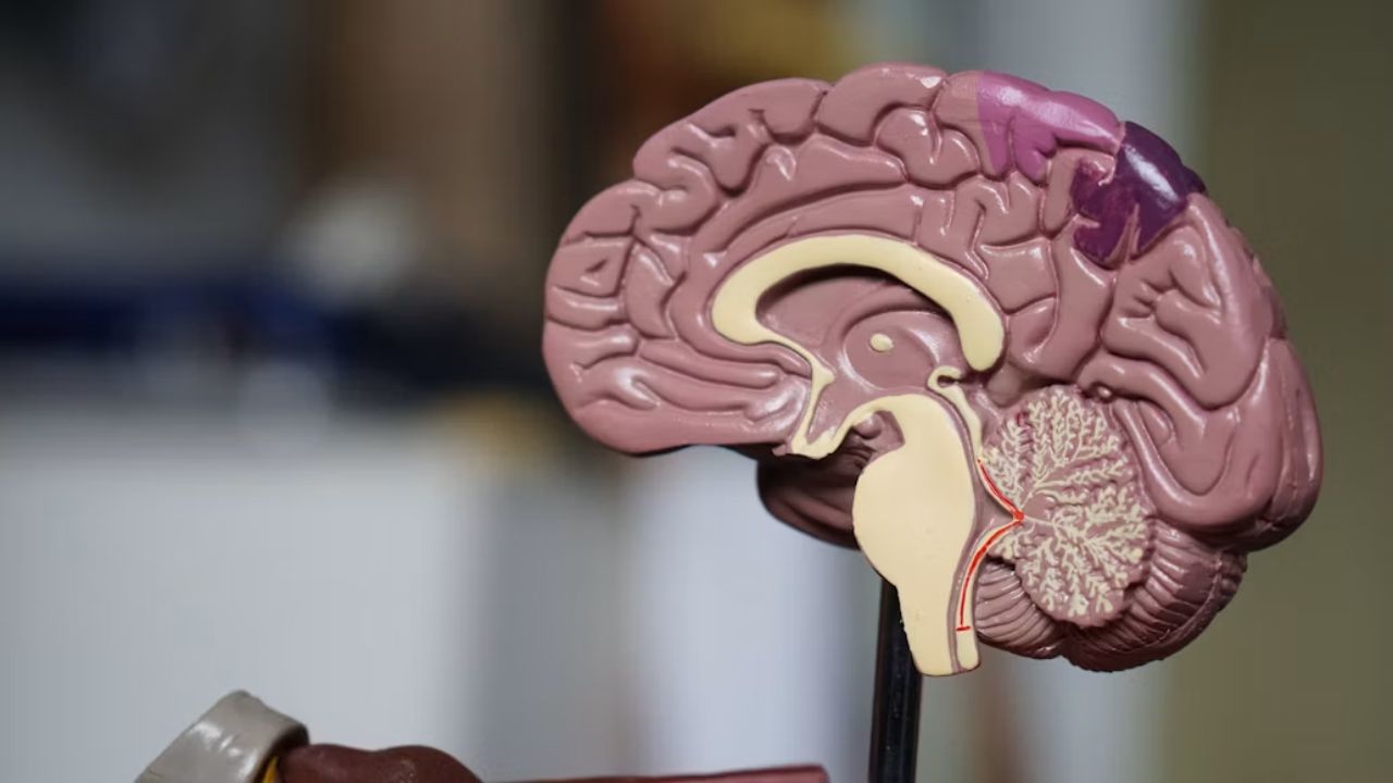
The brain, for long, has been understood as one complex organ made up of several interconnected regions. However, in a new study, researchers found that the brain develops from two distinct groups of early cells, rather than one shared group as previously thought. These two groups eventually give rise to different parts of the brain, with one forming the forebrain and midbrain and the other forming the hindbrain. The findings were published in Nature Neuroscience, and challenge a long-standing belief of how the brain develops.
The researchers say this could mean that the brain is essentially made up of two ancient nervous systems that came together during evolution. This does not mean that humans have two physically separate brains. Instead, the discovery is about how the brain develops and where its different regions come from.
Kyle Loh, PhD, associate professor of developmental biology, and senior author of the study said, "We've shown for the first time that the front of the brain arises from a totally different progenitor cell than the back of the brain," said. "Our discovery means that we can now grow neurons from the back of the brain, the hindbrain, in a petri dish and study their functions." Graduate students Carolyn Dundes and Rayyan Jokhai are co-first authors of the research.
What Did The Stanford Study Find?
The adult brain is generally divided into three major regions: the forebrain, midbrain and hindbrain. These areas have different roles. The forebrain is involved in functions such as language, consciousness and abstract thinking, while the hindbrain, which includes the brainstem, controls several automatic processes needed for survival, including breathing and regulation of the heartbeat. Hindbrain neurons also help control muscles involved in facial movements, speaking and swallowing.
The researchers found that these regions do not all begin from the same type of early cell. Instead, they identified two separate populations of progenitor cells during early embryonic development.
One population, associated with the Otx2 gene, develops into the forebrain and midbrain. The other, associated with Gbx2, develops into the hindbrain. The two populations remained separate and did not overlap in the experiments.
How Does The Brain Develop From Two Cell Groups?
The difference begins very early in embryonic development, during a stage called gastrulation. This is when the basic body plan starts taking shape. The researchers studied developing mouse embryos and used lineage tracing to follow where different groups of cells went. Their findings showed that the cells destined to form the forebrain and midbrain followed one developmental pathway, while cells that would form the hindbrain followed another.
The team also studied chromatin, the material that packages DNA inside cells. Chromatin helps determine which genes a cell can access and use. Researchers found major differences in chromatin configuration between the developing regions that would become the forebrain and midbrain and those destined to become the hindbrain.
This says that the two groups become committed to their respective developmental paths very early, rather than starting as identical cells and gradually becoming different.
Why Scientists Call It Two Separate Organs
The phrase "two separate organs" can sound like the brain is physically divided into two independent structures. That is not what the researchers mean. Instead, the study suggests that the brain has two different developmental origins. The researchers propose that two ancient nervous systems may have evolved separately and were later brought together through evolution.
The idea is supported by experiments involving other animals. Researchers found evidence of similar developmental patterns in chickens, zebrafish and acorn worms, suggesting that the separation may have deep evolutionary roots. The findings therefore give a different way of thinking about the evolution of the brain. What now functions as one connected organ may have originated from two distinct neural systems.
What This Means For ALS And SMA?
In amyotrophic lateral sclerosis (ALS), nerve cells involved in movement progressively become damaged. spinal muscular atrophy (SMA) is a genetic condition in which specific motor neurons are affected. Both conditions can involve neurons associated with swallowing and breathing, making the hindbrain particularly important to researchers studying these diseases.
The finding also helped scientists grow human hindbrain neurons in the laboratory, something that has been difficult to achieve. This could eventually help researchers study conditions such as ALS and SMA, which can damage nerve cells in the brainstem.
The Stanford researchers now want to understand more about how this developmental split occurred and how it relates to the evolution of the nervous system. They also plan to investigate the developmental origins of the spinal cord and study how diseases such as ALS and SMA affect hindbrain neurons.
Disclaimer: This content including advice provides generic information only. It is in no way a substitute for a qualified medical opinion. Always consult a specialist or your own doctor for more information. NDTV does not claim responsibility for this information.